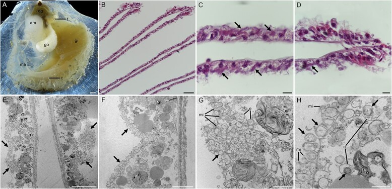
Figure 1.

Anatomy features of Catillopecten margaritatus and photomicrographs showing the extracellular localization of the SOB. A: Anatomy features of C. margaritatus showing the gill tissue, left valve. B-D: Hematoxylin–eosin (HE) stain of gill sections showing a gross view of the gill filaments. E-H: Transmission electron microscopy (TEM) of gill filament showing the extracellular distribution of the SOBs. The bacteria are in the extracellular spaces filled by microvilli, indicated by arrows. Scale bar: A: 1 mm; B: 50 μm; C-D: 10 μm; E: 10 μm; F: 5 μm; G: 2 μm; H: 1 μm. Am, adductor muscle; b: Bacteria; dg, digestive gland; e: Endocytosis; f, foot; go, gonad; gi, gill; ma, mantle; mi: Microvillus; t, tentacle.
