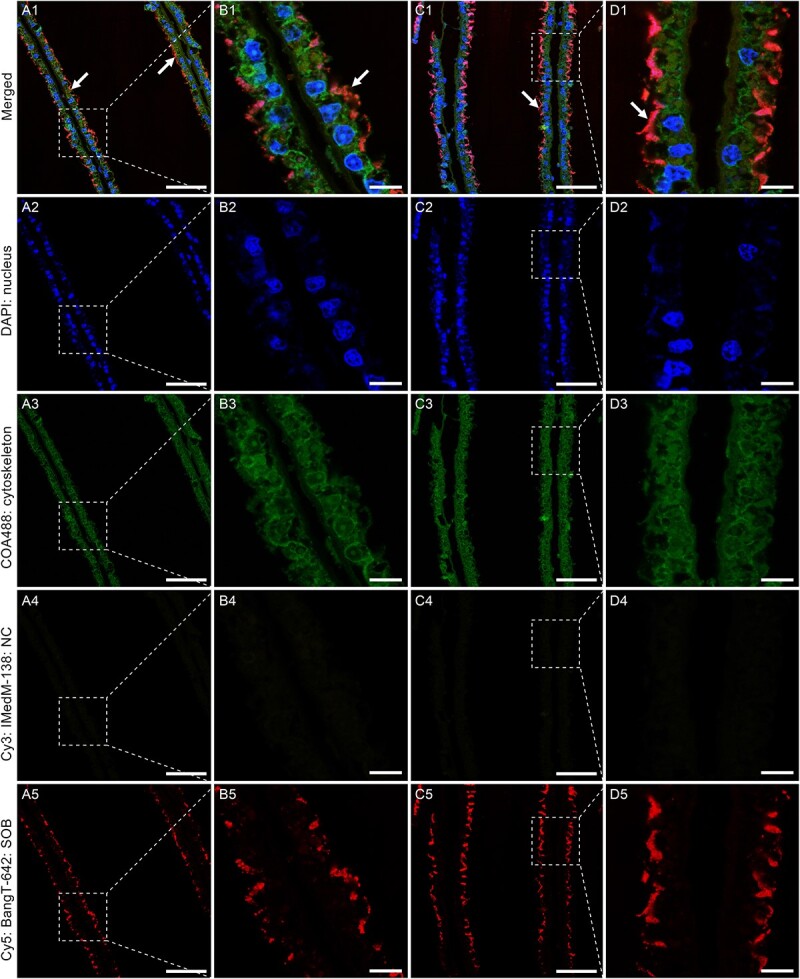
Figure 2.

Fluorescence in situ hybridization (FISH) showing the extracellular localization of the SOB. The DAPI channels show the locations of the nuclei and the COA488 channels represent the gill cytoskeleton. The Cy3 channels show the negative control (NC) using the IMedM-138 probe targeting methanotrophic Gammaproteobacteria. The Cy5 channels indicate the bacteria based on the BangT-642 probe targeting thiotrophic Gammaproteobacteria (SOB). The bacteria are indicated by arrows, and the weak bacterial signals inside the gill cells might be the ectosymbionts endocytosed by the host, as indicated by the TEM micrographs (Fig. 1G and H). Scale bar: A & C: 50 μm; B & D: 10 μm.
