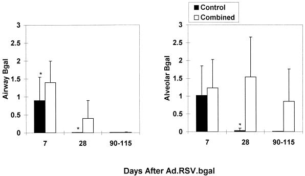
FIG. 2.
The combined-treatment regimen prolongs βgal expression in the lung epithelium. Results are the numerical score for βgal expression based on a scale of 0 to 5 (see Materials and Methods for details of the scoring method). Expression in the bronchial epithelium is shown on the left, and expression in the alveolar epithelium is shown on the right. Data were derived from two to four experiments with 5 to 11 mice for each time point per group and are shown as mean ± standard deviation (SD). ∗, p < 0.05 (control versus combined-treatment group). The combined-treatment group received muCTLA4Ig (200 μg) and MR1 (250 μg) intraperitoneally on days −1, 2, and 7 and Ad.RSV.muCTLA4Ig intratracheally at the time of Ad.RSV.βgal administration. The control group received L6 (200 μg) intraperitoneally on days −1, 2 and 7 and Ad.RSV.hAAT intratracheally at the time of Ad.RSV.βgal administration.

