. 2024 Mar 12;105(3):001964. doi: 10.1099/jgv.0.001964

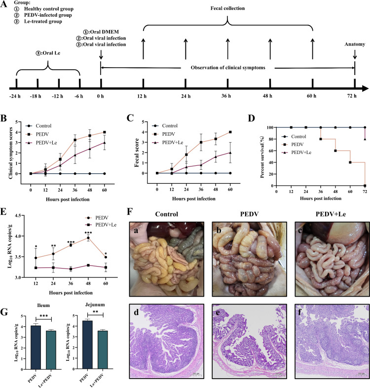
Fig. 5. Oral administration of Le protected piglets against PEDV infection in vivo. (A) Schematic diagram of piglet experiment. (B) Clinical symptom scores of piglets in each group. (C) Faecal scores of piglets in each group. (D) Survival rate of piglets in each group. (E) Detection of faecal virus shedding. (F) Necropsy and histopathology of the intestines of PEDV-infected piglets treated with Le at 72 hpi. Macroscopic pictures of the intestines of the Control, PEDV, and PEDV +Le groups at 72 hpi (a–c). HE-stained of small intestinal tissue sections of piglets in the Control, PEDV, and PEDV +Le groups at 72 hpi (d–f). (G) The distribution of viruses in the jejunum and ileum by qRT-PCR.

Fig. 5.