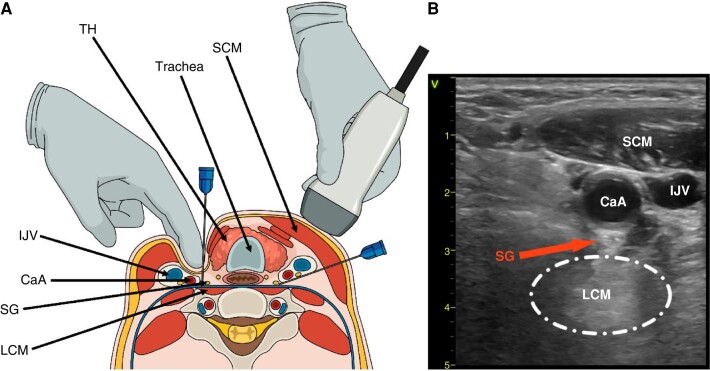
Figure 6.
Stellate ganglion block. (A) Neck cross-section showing SGB guided by surface landmark technique and palpation (left) and ultrasound (right). (B) Transverse sonographic view of the neck at the level of sixth cervical vertebrae (C6). CaA, carotid artery; IJV, internal jugular vein; LCM, longus colli muscle; SCM, sternocleidomastoid muscle; SG, stellate ganglion; SGB, SG blockade; TH, thyroid.

