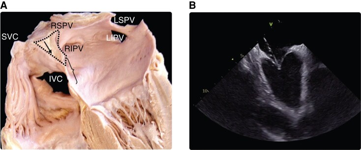
Figure 3.
(A) Anatomy of interatrial septum and optimal site of transseptal puncture (demarcated with a brace). Black arrow in the dotted area shows the infolded groove of the atrial wall between the SVC and the right PVs filled with extracardiac fat tissue. (B) Intracardiac echo view of typical tenting before transseptal crossing. Modified from Tzeis et al.159 IVC, inferior vena cava; LIVP, left inferior pulmonary vein; LSVP, left superior pulmonary vein; PV, pulmonary vein; RIVP, right inferior pulmonary vein; RSVP, right superior pulmonary vein; SVC, superior vena cava

