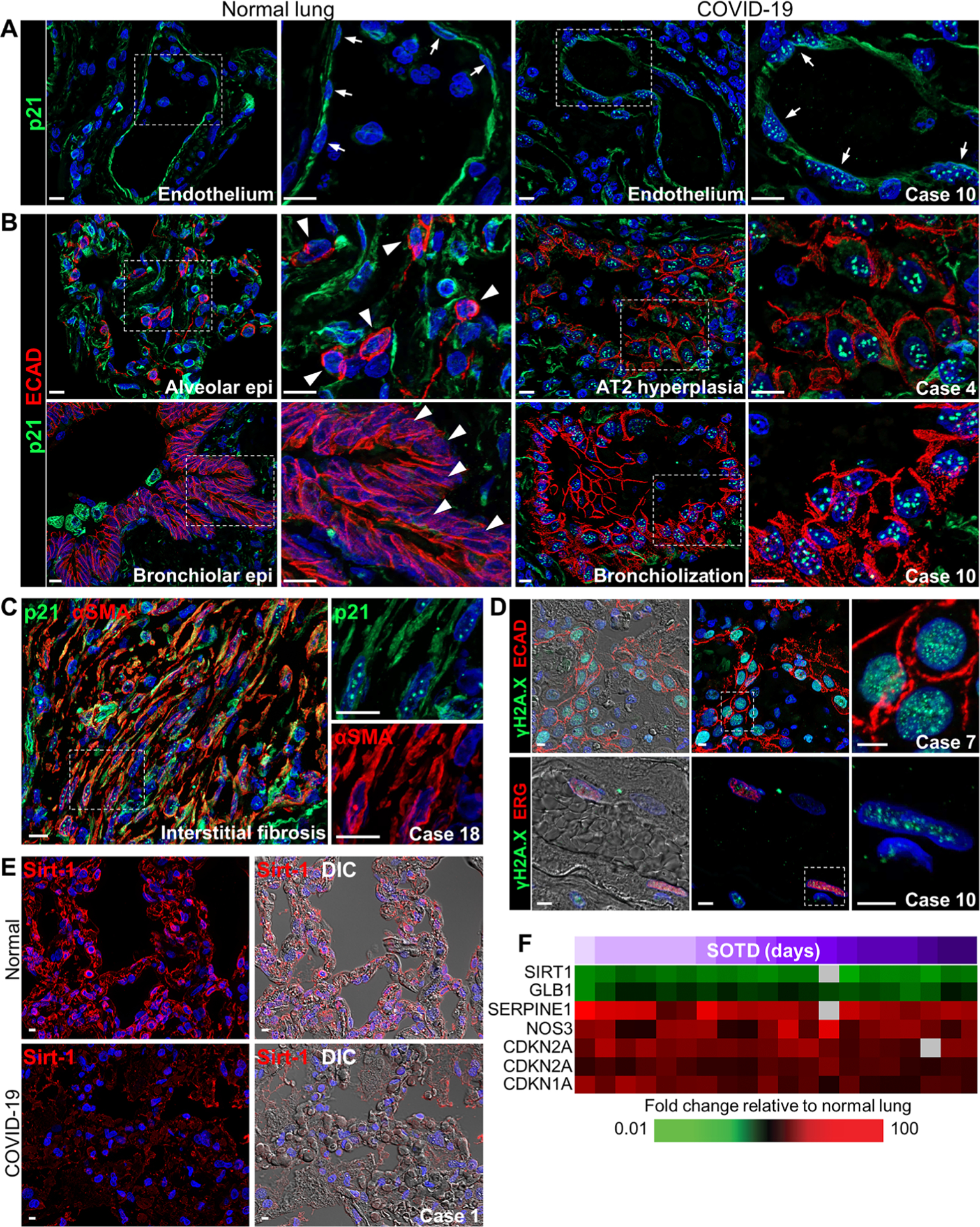
Fig. 7. Expression of cellular senescence markers in COVID-19 lung autopsy samples.

(A and B) Representative images show p21 staining in (A) vascular endothelium and (B) alveolar and airway epithelium in normal lung tissue and COVID-19 lung tissue (cases 4 and 10). (A) Distinctive punctate nuclear p21 staining is observed in endothelial cells in the COVID-19 lung section but not in the normal lung section (white arrows). (B) Punctate p21 nuclear staining is seen in E-cadherin–labeled epithelial cells in abnormal hyperplastic and bronchiolization lesions in COVID-19 lung tissue. White arrowheads depict the lack of nuclear p21 staining in normal lung AT2 cells and bronchiolar epithelial cells. (C) Representative immunofluorescence images show colocalized staining of p21 and α–smooth muscle actin (αSMA) in cells forming interstitial fibrotic lesions in lung tissue from a patient with COVID-19 (case 18) with a longer SOTD. White dotted box shows area of digital enlargement. (D) Immunofluorescence and DIC images show nuclear γH2A.X foci in epithelial (top) and endothelial (bottom) cells costained for E-cadherin or the erythroblast transformation specific (ETS)–related gene (ERG) in COVID-19 lung tissue (cases 7 and 10). (E) Representative immunofluorescence and DIC images show sirtuin-1 (Sirt-1) expression in the alveolar septa of a normal lung section and a COVID-19 lung section (case 1). (F) Heatmap shows expression of genes encoding senescence markers with differential expression in COVID-19 lung tissue samples (n = 13 cases; table S5) compared to normal lung tissue. Time (days) of SOTD is indicated by a purple gradient bar. Scale bars, 10 μm (A to C) and 5 μm (D and E). Epi, epithelium.
