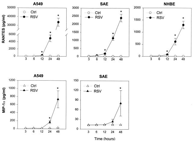
FIG. 3.
Kinetics of RANTES and MIP-1α accumulation in supernatant from RSV-infected A549, SAE, and NHBE cells. Epithelial cells were infected with RSV at an MOI of 1 (RSV) or cultured in control medium (Ctrl). After indicated time of incubation, supernatants were collected for RANTES and MIP-1α determination by ELISA. The results are expressed as mean ± SD of four experiments. ∗, P < 0.05 compared with control at each time point.

