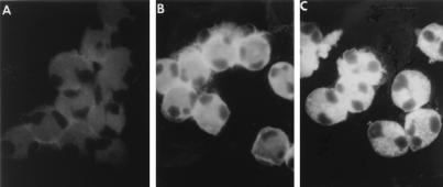
FIG. 7.
Eosinophil infection by RSV. Eosinophils were cultured with control medium (A) or infected with RSV for 2 h (B) or 16 h (C). Cytospin preparations of eosinophils were stained with anti-RSV Fgp MAb followed by a fluorescein isothiocyanate-conjugated anti-mouse F(ab′)2 IgG antibody. In the preparations of eosinophils that were exposed to RSV for 2 h, the Fgp staining was concentrated in a pericytoplasmic halo. Upon RSV infection for 16 h, typical intracytoplasmic granular fluorescence immunoreactivity was observed.

