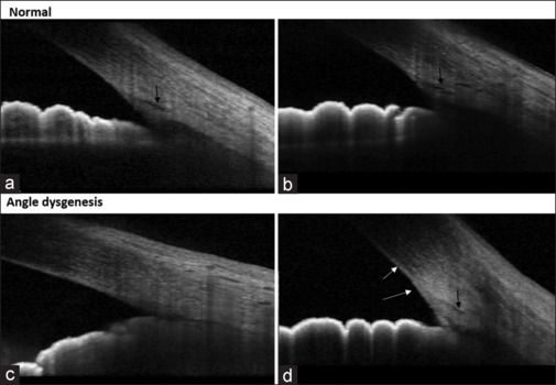
Figure 1.

Representative high-definition AS-OCT images of normal healthy eyes (a and b) versus those with angle dysgenesis (c and d). Black arrows show the location of Schlemm’s canal, and the white arrows show the presence of a hyperreflective membrane at the angle
