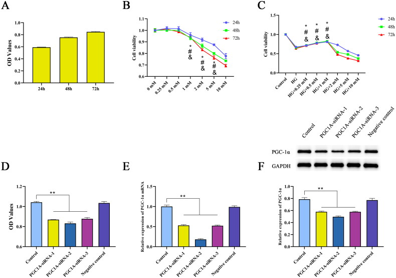
Figure 8.
Concentration screening and siRNA-PGC-1α transfection. Cell viability was evaluated by CCK-8 assay. (A) Cell viability of HK-2 cells incubated with 30 mM glucose for 24, 48, and 72 h. (B) Cell viability of HK-2 cells incubated with NaBut (0.25, 0.5, 1, 2, 5, 10 mM) for 24, 48, and 72 h, *p < 0.01 vs. the 0.5 mM group in 24 h, #p < 0.01 vs. the 0.5 mM group in 48h, &p < 0.01 vs. the 0.5 mM group in 72h. (C) Cell viability of DKD cell model incubated with NaBut (0.25, 0.5, 1, 2, 5, 10 mM) for 24, 48, and 72 h, *p < 0.01 vs. the HG group in 24 h, #p < 0.01 vs. the HG group in 48h, &p < 0.01 vs. the HG group in 72h. (D–F) Cell viability, mRNA and protein expression of PGC-1α following transfection with siRNA-PGC-1α HK-2 cells cultured with 5 mM glucose served as the control for all experiments. **p < 0.01, group sizes = 3.

