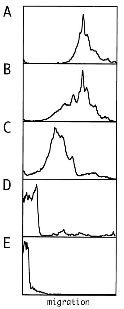
FIG. 3.
Electrophoretic analysis of RNPs assembled in vitro with recombinant δAg-S and in vitro-transcribed genomic HDV RNA. (A) Profile of 500 ng of genomic RNA synthesized in vitro. (B to E) Aliquots of 500 ng of RNA incubated with 67, 200, 600, and 900 ng of δAg-S, respectively. A trace amount of 32P-labeled genomic RNA was included to allow detection following electrophoresis. Quantitation was via a phosphoimager (Fuji). Images were further processed with Canvas software. In each panel, electrophoresis was from left to right, with the gel origin shown at the left side.

