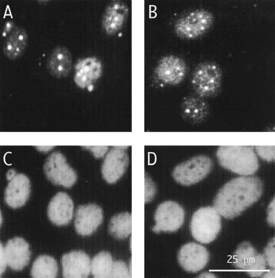
FIG. 5.
Immunofluorescence microscopy to detect genome replication following transfection of cells with assembled RNPs. RNPs were assembled as described in the legend to Fig. 3C. At day 8 after transfection, immunofluorescence was used to detect either total δAg (A) or, specifically, δAg-L (B). (C and D) Same fields as panels A and B, but with detection of cellular DNA by DAPI staining. The scale is indicated in panel D.

