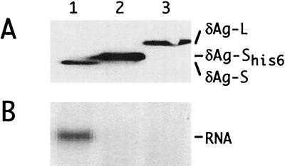
FIG. 8.
Immunoblotting and Northern analyses following transfection of cells with cDNAs. Cells were transfected with pSVL(D2m) together with one of the following vectors: lane 1, pDL444 (δAg-S); lane 2, pTW203 (δAg-SHis6); and lane 3, pDL445 (δAg-L). At 6 days, δAg expression was examined by immunoblotting (A) and HDV genomic RNA was examined by Northern analysis (B). To the right are indicated the different δAg species detected by immunoblotting and the unit-length HDV RNA detected by Northern hybridization.

