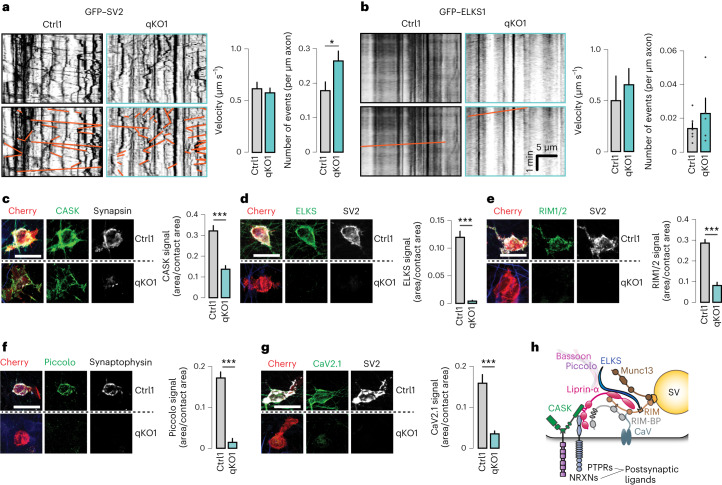
Fig. 4. Removal of liprin-α does not affect axonal transport but prevents accumulation of presynaptic proteins at nascent contacts.
a, The transport of synaptic vesicle cargo in liprin-α control (Ctrl) and mutant (qKO) neurons. Left, representative kymographs depicting GFP-tagged SV2 (GFP–SV2) movements (highlighted in orange in lower images) along axons. Scale as in b. Right, summary plots for the velocity and frequency of transport events. Number of fields(particles)/batches: Ctrl1, 13(310)/2 and qKO1, 14(501)/2. b, The transport of synaptic vesicle cargo in liprin-α Ctrl and qKO synapses. Left, representative kymographs depicting GFP-tagged ELKS (GFP–ELKS) movements (highlighted in orange in lower images) along axons. Right, summary plots for the velocity and frequency of transport events. Number of fields(particles)/batches: Ctrl1, 10(22)/3 and qKO1, 12(22)/3. c–g, The recruitment of CASK (c), ELKS (d), RIM (e), piccolo (f) and CaV2.1 (g) to HEK293 cells expressing Nlgn1. Left, representative images. Right, summary statistics. Number of cells/batches: 79–158/3–4 (Supplementary Table 1). h, A schematic model of a presynaptic terminal highlighting the main active zone components, calcium channels and key presynaptic CAMs, based on known protein–protein interactions6,34. SV, synaptic vesicle. Scale bars, 20 μm. Data are represented as means ± s.e.m. *P < 0.05 and ***P < 0.001.

