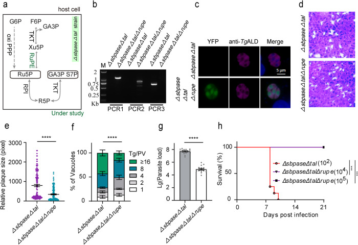
Fig. 3. Deleting RuPE in the ΔsbpaseΔtal mutant harms parasite growth and virulence.
a Scheme of the R5P synthesis pathway in the ΔsbpaseΔtal strain, highlighting the position of RuPE enzyme. b Diagnostic PCRs confirming the ΔsbpaseΔtalΔrupe mutant. c Immunostaining of the ΔsbpaseΔtal and ΔsbpaseΔtalΔrupe (YFP positive) strains with the rabbit anti-TgALD antibody. Scale bars = 5 μm. d Plaque assay assessing the comparative growth of the ΔsbpaseΔtalΔrupe mutant and its progenitor strain in HFF cells. e Plaque size based on the assay in d (n = 3 experiments, means ± SEM, unpaired two-tailed Student’s t-test; ****p < 0.0001). f Replication efficiency as determined by parasite distribution in the parasitophorous vacuoles. The number of parasites/vacuole was counted 24 h post-invasion (means ± SEM of three independent experiments, each with two replicates; ****p < 0.0001, two-way ANOVA). g Parasite burden in the peritoneal fluid of female ICR mice infected by the ΔsbpaseΔtal and ΔsbpaseΔtalΔrupe mutants (104 tachyzoites per mouse). The parasite load was assessed by qPCR after 5 days of infection (5 mice/group; n = 3 assays; ****p < 0.0001, unpaired two-tailed Student’s t-test). h Virulence test in ICR mice infected with a dose of 102 parasites of the ΔsbpaseΔtal strain (8 mice). The ΔsbpaseΔtalΔrupe mutant was also inoculated at higher doses (104, 105 parasites; 5 mice/group). Statistical significance was tested by log rank Mantel–Cox test. Compared with ΔsbpaseΔtal strain (***p = 0.0004). Source data are provided as a Source data file.

