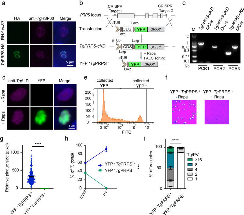
Fig. 7. Knockout of PRPS impairs parasite proliferation.
a Immunolocalization of TgPRPS-HA with HSP60 (a mitochondrial marker, 1:1000). The transgenic strain was generated by CRISPR-assisted 3′-genomic tagging. Scale bars = 5 μm. b Replacement of PRPS in the DiCre strain by CRISPR/Cas9-mediated homologous recombination. c Diagnostic PCRs on a conditional mutant (TgPRPS-cKD). d Immunofluorescence staining of TgALD and expression of YFP in the TgPRPS-cKD mutant incubated with rapamycin for 5 days. Scale bars = 5 μm. e Flow cytometry of YFP expression in rapamycin-treated mutant. f, g Plaque assay of the TgPRPS-cKD strain (−/+ rapamycin, 5 days). (n = 3 experiments, means ± SEM, ****p < 0.0001, unpaired two-tailed Student’s t-test). h Competition assay (−/+ rapamycin, 5 days) (n = 3 experiments; means ± SEM; P1, ***p = 0.0002, unpaired two-tailed Student’s t-test). i Replication rates of the TgPRPS-cKD mutant (−/+ rapamycin, 5 days). Parasites expressing YFP (YFP+TgPRPS−) were sorted by FACS. Intracellular tachyzoites (24 h infection) replicating in parasitophorous vacuoles were counted (Tg/PV; n = 3 assays, means ± SEM; ****p < 0.0001, two-way ANOVA). Source data are provided as a Source data file.

