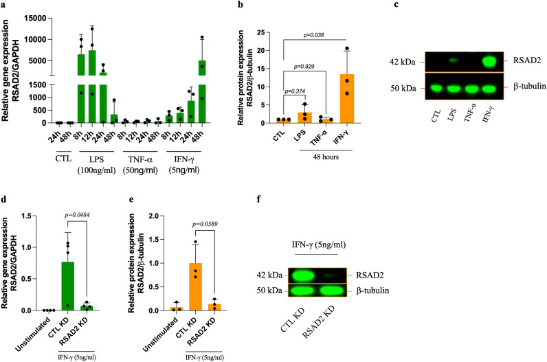
Figure 2.
Inflammatory mediators induce RSAD2 in human aortic smooth muscle cells and RSAD2-siRNA efficiently reduces IFN-γ-induced RSAD2 expression. (a) Relative mRNA expression of RSAD2 in hAoSMCs stimulated with IFN-γ (5ng/ml), LPS (100µg/ml) or TNF-α (50ng/ml) for 8–48 h (n = 3). (b) Relative protein expression of RSAD2 in hAoSMCs exposed to LPS (100µg/ml), TNF-α (50ng/ml) or IFN-γ (5ng/ml) for 48 h (n = 3). (c) Representative cropped Western blot image of RSAD2 and β-tubulin from hAoSMCs exposed to LPS, TNF-α or IFN-γ for 48 h (full-length Western Blot images are shown in supplementary fig. S2a). (d) Relative mRNA (n = 4) and (e) protein (n = 3) expression of RSAD2 in hAoSMCs transfected with 30pmol of RSAD2-targeting or scramble siRNAs and stimulated with IFN-γ for 44 h (normalized to the average of CTL KD). (f) Representative cropped Western Blot image of RSAD2 and β-tubulin from IFN-γ-stimulated hAoSMCs transfected with RSAD2-targeting or scramble siRNAs (full-length Western Blot images are shown in Supplementary Fig. S3a). The data are presented as mean ± SD. One-way ANOVA followed by Bonferroni's multiple comparison test was used to evaluate statistical significance. p value < 0.05 is considered statistically significant. LPS, lipopolysaccharide; TNF-α, tumor necrosis factor alpha; IFN-γ, interferon gamma; RSAD2, radical S-adenosyl-L-methionine domaine containing 2.

