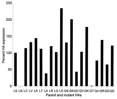
FIG. 1.
Cell surface expression of mutant HAs analyzed by FACS. Cos-1 cells (60% confluency) were transfected with 2 μg of purified plasmid DNA per well of a 6-well tissue culture plate with Lipofectamine (Gibco). The cells and transfection mixture were incubated for 5 h at 37°C, after which the transfection medium was replaced with 2 ml of Opti-Mem medium (Life Technologies, Inc.) containing 5% fetal calf serum (FCS). The cells were then incubated for 40 h at 37°C. Cells expressing HA were washed with PBS, treated with Vibrio cholerae sialidase (5.5 milliunits/ml; Life Technologies, Inc.) for 1 h at 37°C (which abolishes the susceptibility of cells to influenza virus infection) to remove sialic acid from the HA, which interferes with receptor recognition (17), and then maintained in suspension following trypsinization. Cells were spun down, washed twice with PBS containing 10% FCS, and then resuspended in PBS containing 10% FCS and an anti-H3 HA monoclonal antibody (S11/4, S28/1, S37/2, and 121/1) pool (diluted 1:400). After a 1-h incubation at 4°C, the cells were washed twice with PBS containing 10% FCS, and then incubated with fluorescein isothiocynate-labeled goat anti-mouse immunoglobulin (diluted 1:20 in PBS containing 10% FCS) (Boehringer Mannheim Biochemicals) for 30 min at 4°C. The cells were washed twice as before and then fixed in PBS containing 3.7% paraformaldehyde for 20 min at 4°C. Cells were centrifuged, resuspended in PBS containing 10% FCS, and stored at 4°C until analyzed by FACS with a FACScan (Becton Dickinson). The relative expression levels presented are based on mean values of fluorescence intensity. Experiments were repeated three times, and representative data are shown.

