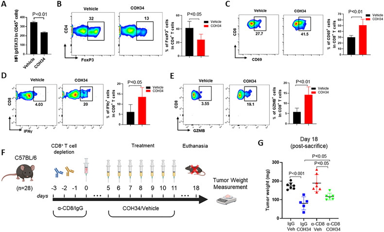
Figure 5.
In vivo PARG inhibition induces antitumor immune responses, and the antitumor effects are partially mediated by CD8+ T cells. 5×10⁶ Brca1-null ID8 tumor cells were injected subcutaneously into female C57BL/6 mice aged 7–10 weeks old. The tumor-bearing mice were treated with vehicle or COH34 (20 mg/kg) every day for 7 days. Single-cell suspensions prepared from the tumors were analyzed by flow cytometry to detect: (A) phosphorylated STAT3 (pSTAT3) in CD45+ immune cells. (B) FoxP3+ Tregs in CD4+ T cells and; (C, D, E) activated CD8+ T cells (CD69+ cells, IFN-γ+, and GZMB+). Data are shown as means±SD (n=4–5, each sample was pooled from 3 to 4 mice). (F) In vivo study design for (G). To deplete CD8+ T cells, C57BL/6 mice were injected intraperitoneally with rat anti-CD8 antibody or rat IgG2b (isotype control) on days −3 to –2, −1, and 0 relative to subcutaneous injection of Brca1-null ID8 tumor cells (day 0). When the tumors reached an average size of 100 mm³ on day 5, vehicle or COH34 (20 mg/kg) were administered by intraperitoneal injections every day for 7 days. (G) On day 18, mice were euthanized and tumor weight was measured. Data are shown as means±SD (n=5–7 mice per group). PARG, poly(ADP-ribose) glycohydrolase; STAT3, signal transducer and activator of transcription 3.

