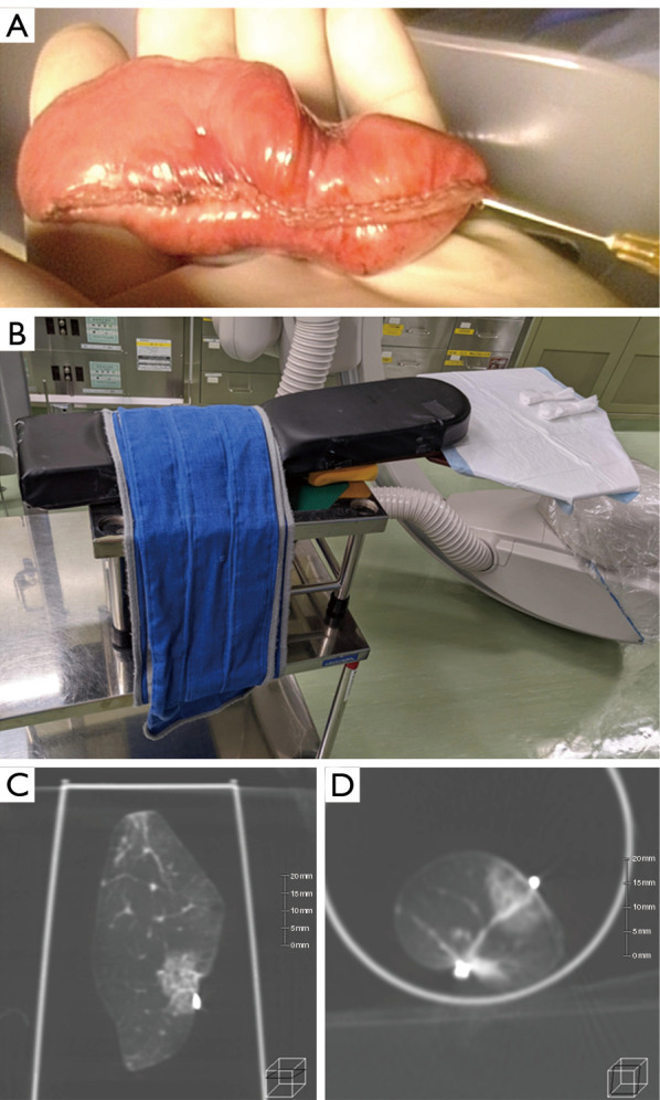
Figure 3.

Resected-lung-scan images. (A) Air injection is performed using a low-gauge needle before the Resected-lung-scan. Note that rapid air injection causes emphysema in the resected lung. (B) A platform for Resected-lung-scan assembly using the arm board of an operating table. A plastic cup containing the specimen is placed on this platform for Resected-lung-scan imaging. (C,D) A Resected-lung-scan indicates the relationship of the lesion to the clips and staple line, and confirms that the resection margin is adequate.
