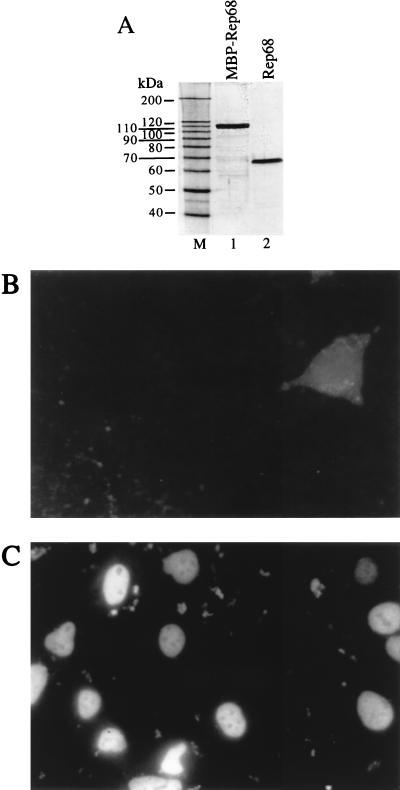
FIG. 2.
Lipofection of Rep68 protein. (A) Silver staining of a sodium dodecyl sulfate-polyacrylamide gel. Rep68 protein was expressed in E. coli as a fusion protein with maltose binding protein (MBP) as described elsewhere (4) and was partially purified by amylose affinity chromatography (lane 1), cleaved with Factor Xa to remove the maltose-binding moiety, and purified to homogeneity (lane 2) by fast protein liquid chromatography with MonoQ (anion exchange) and Superdex-75 (gel filtration) columns (both from Pharmacia). M, molecular size markers. (B and C) Intracellular localization of lipofected Rep68 protein. Two hours after transfection, 293 cells were washed, fixed in 3% formaldehyde, and permeabilized by treatment with 0.1% Triton X-100. The intracellular location of Rep68 was monitored by sequential incubation with the mouse monoclonal anti-Rep antibody 226.7 (Progen) and a rhodamine-conjugated anti-mouse immunoglobulin G goat polyclonal serum. Shown are results of staining of cells incubated for 2 h with Rep68 alone (B) or with the Rep68–Lipofectamine complex (C).

