Fig 3.

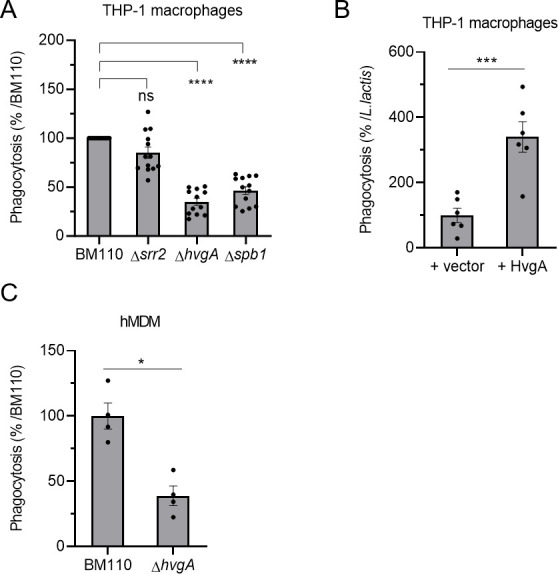
The HvgA surface protein and Spb1 pilus protein are involved in CC17 hyper-phagocytosis. (A–C) Phagocytosis level of GBS strains was assessed by CFU count after infection at MOI 10 followed by antibiotic treatment to kill extracellular bacteria using (A, B) THP-1 macrophages or (C) hMDM primary cells. (A, C) Macrophages were infected with BM110 and its derivative mutant strains (Δsrr2, ΔhvgA, or Δspb1) or (B) with L. lactis strain carrying an empty vector (+vector) or a vector expressing the HvgA protein (+HvgA). (A, C) Results are expressed as the relative levels to BM110 strain or (B) to the control L. lactis strain +vector. Statistical analysis: data shown are mean ± SEM of at least four independent experiments. (A) Kruskall Wallis test with Dunn’s multiple comparisons or (B, C) t test was performed with ns, non-significant; *P < 0.05; ***P < 0.001; ****P < 0.0001.
