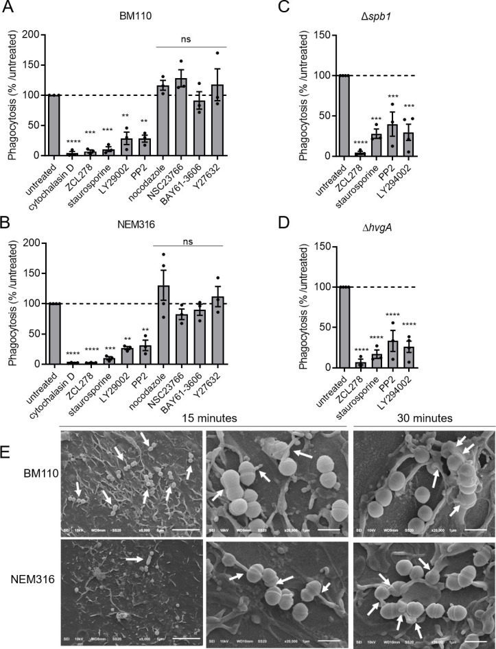
Fig 4.

Signal transduction leading to phagocytosis is similar for CC17 and non-CC17 strains. (A–D) THP-1 macrophages were pre-treated for through D) THP-1 macrophages were pre-treated for 1-h prior to infection with cytochalasin D, ZCL178, staurosporin, LY29002, PP2, nocodazole, NSC23766, BAY61-3606, or Y27632 to inhibit actin polymerization, CDC42, protein kinase C, PI3kinase, Src kinase, microtubule polymerization, Rac, Syc, and Rock, respectively. (A) Phagocytosis level of BM110, (B) NEM316, (C) Δspb1, or (D) ΔhvgA GBS strains was assessed by CFU count after infection at MOI 10 followed by antibiotic treatment to kill extracellular bacteria. Results are expressed as the relative levels to untreated condition. (E) SEM micrographs of THP-1 macrophages showing (left panel, scale bar 5 µm) bacterial adherence to cell surface and (middle panel and right panels, scale bar 1 µm) bacterial capture by filopodia after 15 min or lamellipodia after 30 min of infection. Statistical analysis: data shown are mean ± SEM of at least four independent experiments. One-way ANOVA with Dunnett multiple comparisons was performed with ns, non-significant; **P < 0.01; ***P < 0.001, ****P < 0.0001.
