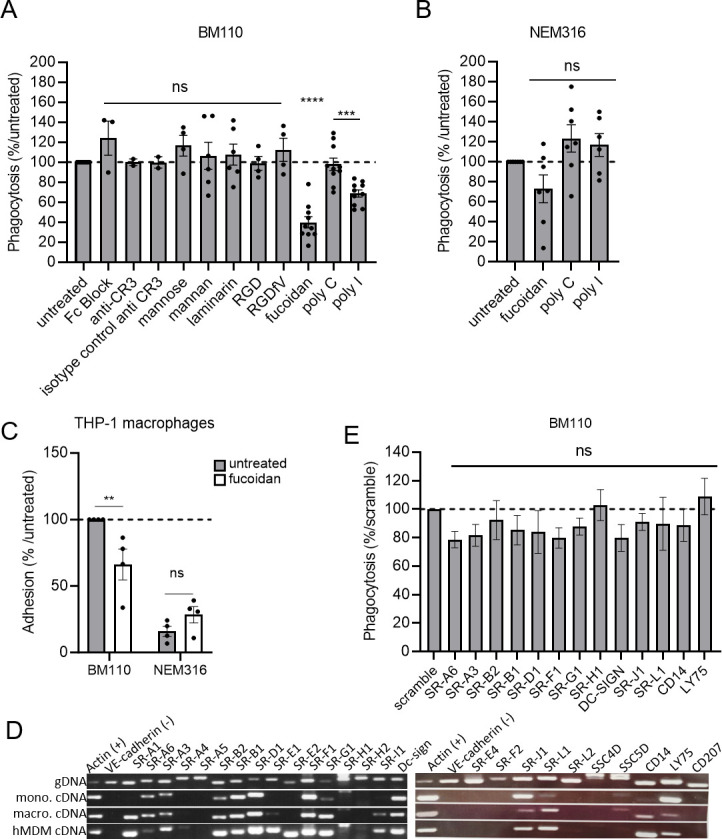
Fig 6.

Scavenger receptor inhibitors lead to a decreased phagocytosis level of CC17 GBS in macrophages. (A, B, E) Phagocytosis level in THP-1 macrophages was assessed by CFU count after infection at MOI 10 with (A, E) BM110 or (B) NEM 316 GBS strains followed by 1 h antibiotic treatment to kill extracellular bacteria. (A) Macrophages were pre-treated 1 h before infection to block FCγ receptors (Fc Block), complement receptor 3 (CR3 blocking antibody), lectin receptors (mannose, mannan, laminarin), integrins receptors (RGD and RGDfV mimetic peptides), or (A, B) scavenger receptors inhibitors [Fucoidan and poly(I) or inactive analog poly(C)]. (A, B) Results are expressed as the percentage of phagocytosis relative to the untreated control condition except for poly(I) that was relative to its inactive analog poly(C). (C) Adhesion level in untreated or fucoidan treated-THP-1 macrophages was assessed by CFU count after infection with BM110 and NEM316 at 4°C to avoid bacterial engulfment. Results are expressed as the percentage of phagocytosis relative to the untreated control condition. (D) Expression of scavenger receptors genes was assessed by RT-PCR using THP-1 macrophages (macro), THP-1 monocyte (mono), and hMDM cDNA. Actin and VE-cadherin gene expression was used as positive and negative controls of expression, respectively. Genomic DNA (gDNA) was used to validate primers efficiency. (E) The involvement of scavenger receptors on BM110 phagocytosis was assessed by silencing scavenger receptors expression by si-RNA. Results are expressed as the percentage of phagocytosis relative to the scramble control condition. Statistical analysis: data shown are mean ± SEM of at least four independent experiments. (A, B, E) Kruskall Wallis test with Dunn’s multiple comparisons or (C) t test was performed, with ns, non-significant; **P < 0.01; ***P < 0.001; ****P < 0.0001.
