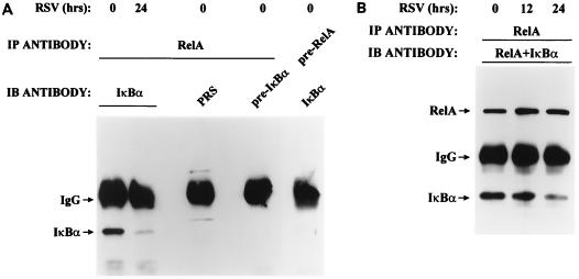
FIG. 3.
Effect of pRSV infection on RelA-associated IκBα. (A) Determination of coimmunoprecipitation assay specificity. RSV-infected (0 or 24 h) WCEs were immunoprecipitated (IP) either with anti-RelA antibody or with preadsorbed RelA antibody (pre-RelA), and the immunoprecipitate was used for the detection of IκBα by Western blotting (IB) using anti-IκBα antibody, preimmune serum (PRS), or preadsorbed IκBα antibody (pre-IκBα). IgG, immunoglobulin G. (B) Degradation of RelA-associated IκBα by RSV infection. Uninfected or pRSV-infected A549 WCE (for 0, 12, or 24 h) was immunoprecipitated with RelA antibody. The immunoprecipitate was subjected to Western immunoblotting with both RelA and IκBα antibodies (Materials and Methods). After 24 h of pRSV infection, RelA-associated IκBα was significantly decreased. For the different time points, the IκBα/RelA ratio was 1.63 (0 h), 1.2 (12 h), and 0.9 (24 h), indicating that IκBα association with RelA falls following pRSV infection.

