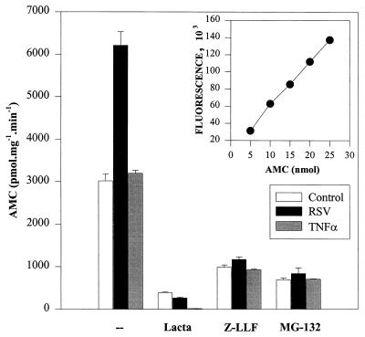
FIG. 7.
26S proteasome activity after treatment and in the presence of proteasome inhibitors. Control, pRSV-infected, or rhTNF-α-treated A549 cells were incubated in the absence or presence of 10 μM lactacystin (Lacta), 10 μM Z-LLF-CHO (Z-LLF), or 25 μM MG-132 for 4 h. After 3.5 h of preincubation, cells were stimulated with rhTNF-α for 0.5 h. Proteasome activities were measured in the cytoplasmic extracts, using the fluorogenic substrate Suc-Leu-Leu-Val-Tyr-AMC (37). Results are expressed as the amount of AMC formed by the enzymatic cleavage of substrate. The inset represents a standard curve for AMC drawn by measuring the fluorescence of known quantities of AMC.

