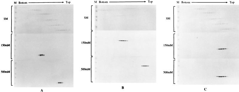
FIG. 2.
Sedimentation profiles of purified Gag antigens on glycerol gradients. Purified soluble Gag antigens were sedimented through 15 to 30% glycerol gradients made in 20 mM Tris (pH 7.4)–100 mM NaCl–1 mM dithiothreitol–0.5 mM EDTA at 48,000 rpm in an SW50 rotor and 4°C for 40 h (for analysis of MA and CA) or for 27 h (for analysis of MA-CA). Molecular mass markers in the gradients were provided by high (analysis of MA-CA)- or low (analysis of MA and CA)-molecular-mass calibration kits (Pharmacia), are shown in the upper section of each panel in all cases, and are marked SM (sedimentation markers). The high-molecular-mass range consisted of the following: catalase, 4 × 58 kDa = 232 kDa; lactate dehydrogenase, 4 × 36 kDa = 140 kDa; and serum albumin, 67 kDa. The low-molecular-mass range consisted of the following: phosphorylase b, 94 kDa; serum albumin, 67 kDa; ovalbumin, 43 kDa; carbonic anhydrase, 30 kDa; trypsin inhibitor, 20.1 kDa; and a-lactalbumin, 14.4 kDa. Gel markers (M) are prestained molecular mass markers (Bio-Rad). Gradients were fractionated from the bottom, and Gag antigen was detected by sodium dodecyl sulfate-polyacrylamide gel electrophoresis and Western blotting. The middle and bottom sections of each panel show an analysis of antigen prepared under low- and high-salt conditions, respectively. (A) MA protein; (B) MA-CA protein; (C) CA protein.

