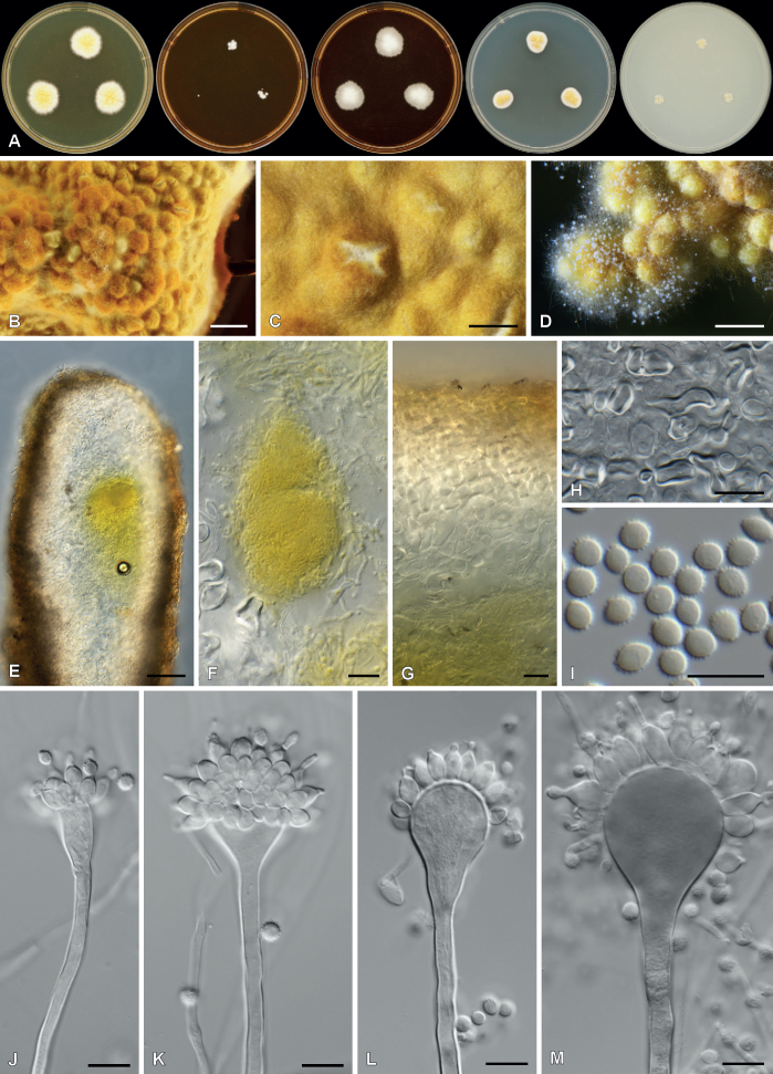
Fig. 35.
Aspergillus lentisci in culture. A. Colonies on, from left to right, MY50G, MY20, MY40, DG18 and OA. B–D. Colony overview on DG18 showing sclerotia and conidiophores (D). E, F. Section of sclerotia showing potential early development of asci and ascomata. G. Section of sclerotia showing its wall structure. H. Hülle cells from sclerotia. I. Conidia. J–M. Conidiophores. Scale bars: B = 2 mm; C, D = 500 µm; E = 50 µm; F–H = 20 µm; I–M = 10 µm.

