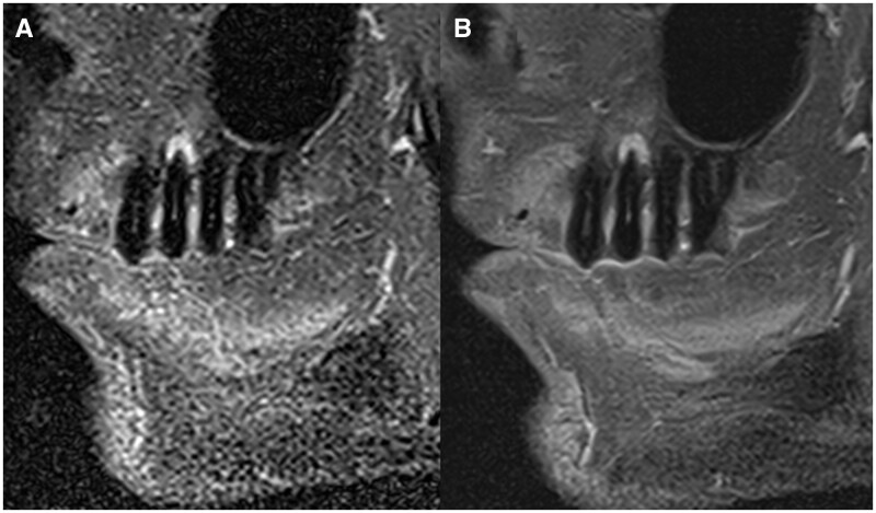
Figure 2.
Comparison of images acquired in the sagittal orientation in a subject with an apical lesion in tooth 25. Both images were acquired with pulse sequences sensitive to inflammatory changes in the bone (ie, fat suppression). In (A), the image was obtained with conventional image acquisition techniques, and in (B), the image was reconstructed using a deep learning-based image algorithm that results in denoized images.

