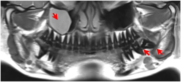
Figure 7.
ddMRI panoramic view, reconstructed from a 3D volumetric acquisition. Presence of a mucous retention cyst in the right maxillary sinus, and a semiimpacted lower third molar in the left side, in which extensive bone loss distal to the second molar and a close relationship between the roots of the tooth and the mandibular canal are shown (red arrows). ddMRI = dental-dedicated MRI.

