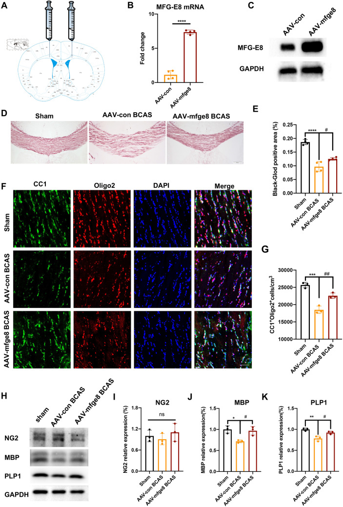
Fig. 4.
Overexpression of MFG-E8 alleviates white matter damage and promotes oligodendrocyte maturation. A Diagram of pAAV-CMV-Mfge8-3xFlag-P2A-mNeonGreen-tWPA injection into the CC. B PCR analysis of MFG-E8 expression in the CC at 3 weeks after stereotaxic intracranial injection (n = 4 per group; ****P < 0.0001, unpaired t-test). C Representative Western blots of MFG-E8 expression in the CC at 3 weeks after stereotaxic intracranial injection. D Representative images of black-gold staining (lavender) in the CC at day 60 after BCAS. Scale bar: 200 μm. E Immunofluorescent intensity of the black-gold staining area in the CC (n = 4, ANOVA; ****P < 0.0001 vs WT sham, #P < 0.05 vs AAV-con BCAS. F Representative immunostaining of CC1 and Olig2 in the CC in the WT sham, AAV-con BCAS at 2 months, and AAV-mfge8 BCAS at 2 months after surgery. Nuclei are stained with DAPI. Scale bar: 50 μm. G Density of CC1+Olig2+ cells in the CC in the above three groups (n = 3 per group; ***P < 0.001 vs WT sham, ##P < 0.01 vs AAV-con BCAS, ANOVA). H Western blots showing the expression of NG2, MBP, and PLP1 in the CC with GAPDH as a loading control. D Densitometry analyses of NG2, MBP, and PLP1 expression normalized to GAPDH (n = 3 per group; *P < 0.05, **P < 0.01 vs WT sham, #P < 0.05 vs AAV-con BCAS, ANOVA. All data are presented as the mean ± SD).

