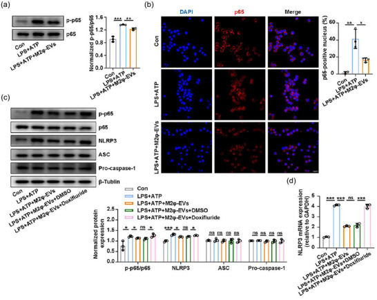
FIGURE 4.

M2φ‐EVs inhibited the NF‐κB/NLRP3 inflammasome pathway. (a and b) After stimulation with LPS (0.5 µg/mL) for 30 min, MH‐S cells were treated with M2φ‐EVs (10 µg/mL) for 23 h and then subjected to ATP (0.5 mM) for another 30 min. (a) Western blotting analysis for phosphorylation levels of p65 in MH‐S cells. The histogram showed the relative ratio of the expression levels of the target protein to that of an internal reference. (b) Nuclear localisation of p65 was visualised by immunofluorescence analysis in MH‐S cells. The histogram represented the percentage of the p65 positive nucleus. (c and d) Pretreated with Doxifluride (10 mM) for 1 h, MH‐S cells were then stimulated with LPS (0.5 µg/mL) for 30 min and then treated with M2φ‐EVs (10 µg/mL) for 23 h and then subjected to ATP (0.5 mM) for another 30 min. (c) Western blotting analysis of p65, p‐p65, NLRP3, ASC and Pro‐caspase‐1 levels in MH‐S cells. The histogram showed the relative ratio of the expression levels of the target protein to that of an internal reference. (d) The mRNA expression of NLRP3 in MH‐S cells was analysed by qPCR. Representative results from three independent experiments are shown (n = 3). Scale bar, 20 µm. ns, not significant, *p < 0.05, **p < 0.01, ***p < 0.001 (one‐way ANOVA test; mean ± SD).
