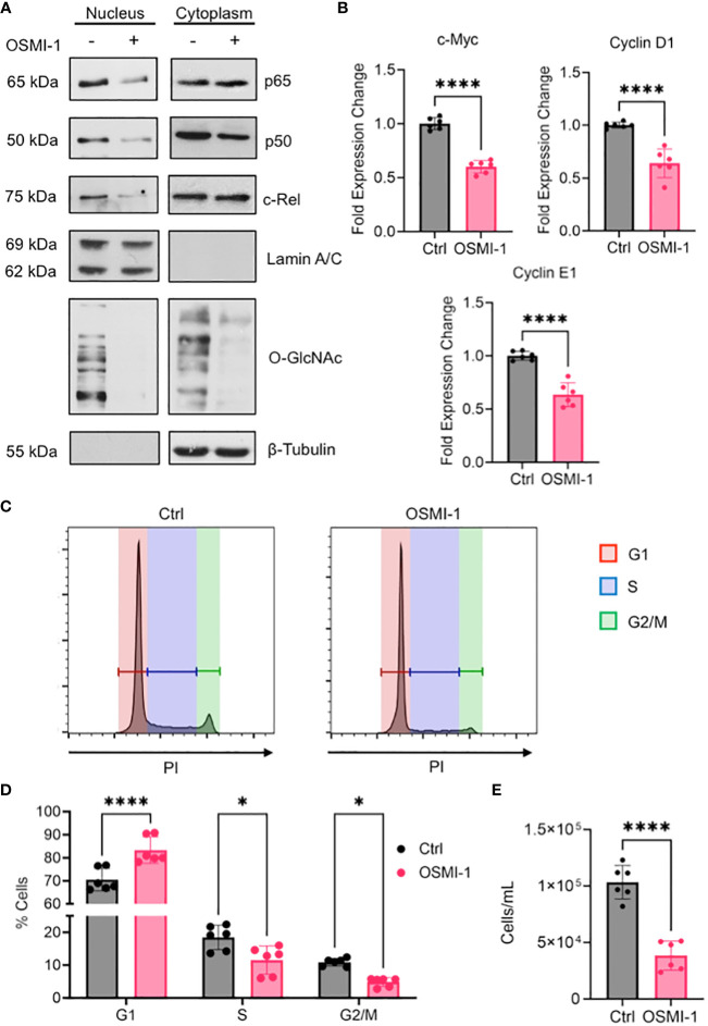
Figure 7.
(A) OCI-AML3 cells were cultured in the presence or absence of OSMI-1 (25 μM) for 4 hours. Cytoplasmic and nuclear extracts were immunoblotted for indicated NF-κB subunits. (B) Gene expression of c-Myc, Cyclin D1, and Cyclin E1 in OCI-AML3 cells after 4 hours of OSMI-1 (25 μM) treatment measured by quantitative real-time PCR. Data shows 3 biological replicates performed with technical duplicates (n = 6). (C) OCI-AML3 cells were treated with OSMI-1 (25 μM) for 48 hours prior to permeabilization and staining with propidium iodide (PI). Representative histograms showing proportion of cells in each phase of the cell cycle: Red=G1, Blue=S, and Green= G2/M. (D) Bar graph showing cell cycle phases representing 3 individual experiments performed with technical duplicates (n = 6). (E) Quantification of cell numbers using flow cytometry after 48 hours of OSMI-1 (25 μM) treatment (n=6). *, P < 0.05; ****, P < 0.0001.

