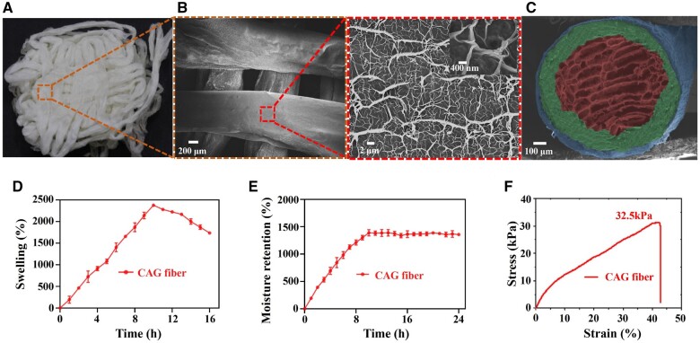
Figure 2.
Microscopic structure and physical properties of CAG fibers. (A) Physical photograph and (B) SEM images of CAG fibers, and the surface of CAG fiber showing a disordered dendritic network structure. (C) SEM image showing the cross-section of CAG fiber with a triple-layered core-shell structure (each layer was pseudo-colored for contrast). (D) Swelling ratio (37°C, pH = 7.4), (E) moisture retention rate and (F) tensile fracture curve of CAG fibers.

