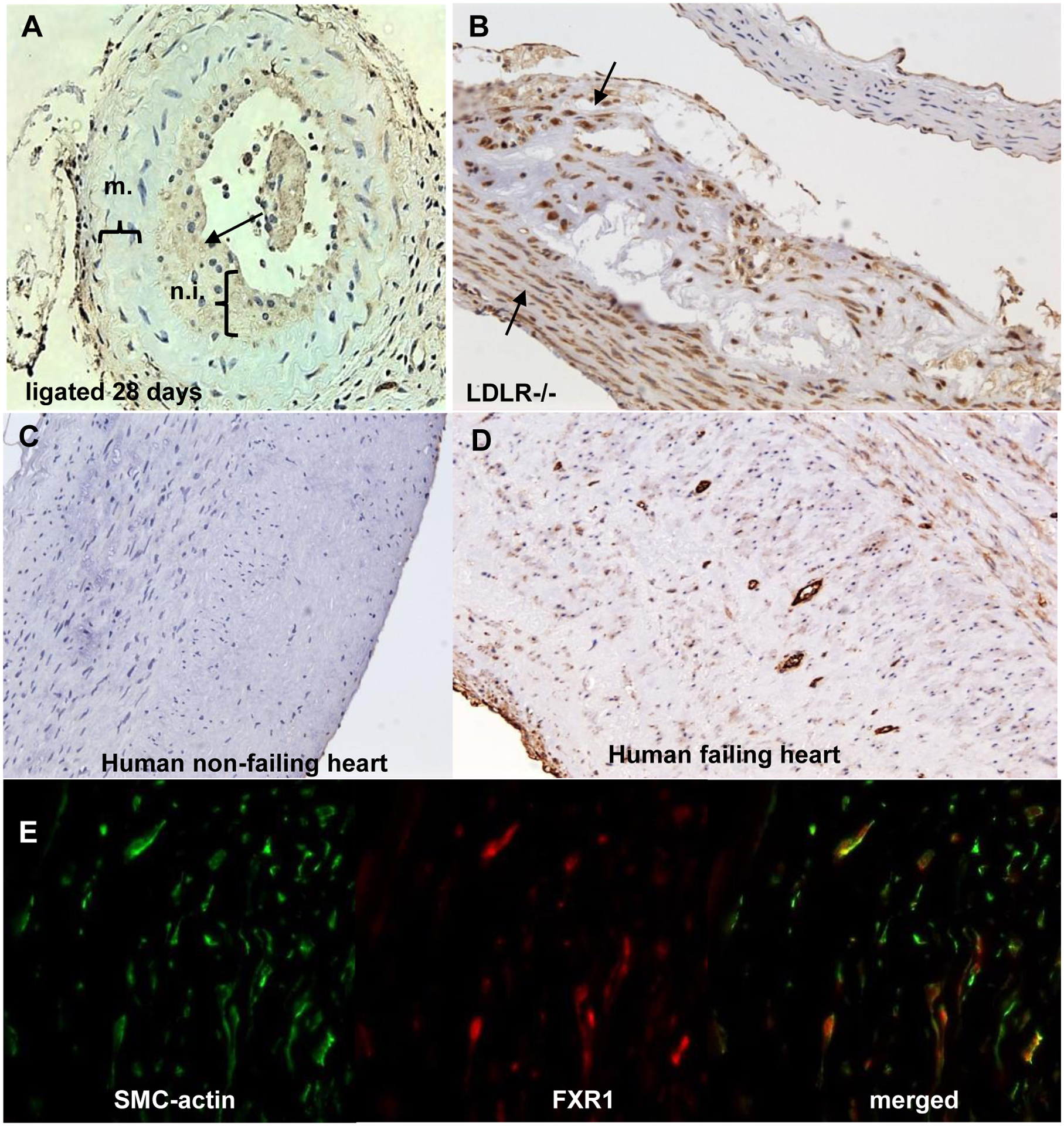
Figure 2.

FXR1 protein expression in vascular injury and disease models. A. FXR1 expression in ligated murine carotid artery. Mouse carotid arteries were harvested 28 days after ligation, and immunohistochemistry performed. FXR1 primarily stains in the neointima (n.i.), but not the media (m.) in the ligated artery. B. FXR1 expression in mouse atherosclerotic plaque. Cross section from an LDLR−/− mouse aorta fed a HFD for 12 weeks to develop atherosclerotic plaque. VSMC in plaque and smooth muscle cell cap are enriched for FXR1 expression. C. FXR1 expression in normal human coronary artery from a non-failing heart. D. FXR1 expression in myleofibroid atherosclerotic plaque from a failing human artery. FXR1 expression is enriched in myleofibroid VSMC in the plaque as compared to healthy human control artery. E. Fluorescent co-staining of fibro-atherosclerotic cap from human atherosclerotic plaque using antibody to SMC-actin and FXR1. See also Supplemental Figure 1 for normal mouse artery and negative controls for immunohistochemistry. Magnification is 200X for all.
