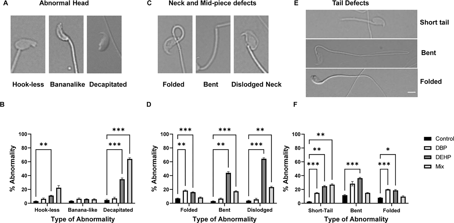
Figure-3: Compartmentalization of sperm morphological defects in the different exposure groups.

Sperm used for the analyses described in figure 2 were further analyzed regarding the type of morphological defects in the head (A and B), mid-piece (C and D) and tail (E and F). Notice that the tail defects do not include double tails, in some of the insets in E, there is superposition of a second spermatozoon’s tail. Spermatozoa with multiple abnormalities were included based on the defects exhibited for each compartment. Data are represented as mean percentage of abnormal sperm ± SEM (N=6). A two-way Anova followed by Dunnett post-hoc test was performed for each compartment of the exposure group with p<0.05 (*), p<0.01 (**), p< 0.001 (***). Data were compared to the control group only.
