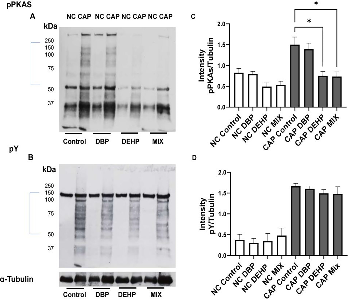
Figure-4: Analysis of sperm capacitation phosphorylation pathways in sperm derived from mice exposed to different phthalate conditions.

A: Phosphorylation of PKA substrates were evaluated by Western blots using anti phospho PKA substrate antibody (pPKAs). Each lane contain protein extracts from ~106 sperm as described in Methods. B: Changes in protein tyrosine phosphorylation were assessed using anti PY antibodies in the same blots used for A after stripping the immobilon membrane as described in Methods. The same blots were stripped once more and probed with anti-tubulin antibody. C and D: Densitometric analyses of pPKAs and PY were done with IMAGE J software. For this analysis, only the section marked with a bar in A and B were used for the evaluation. Pixels were normalized with the values obtained from the anti-tubulin Western blots. Kruskal Wallis non-parametric tests followed by Dunn’s test were performed using GraphPad prism. Data are represented as mean percentage of abnormal sperm ± SEM with p<0.05(*) (N=12),. NC denotes non-capacitated while CAP denotes capacitated condition
