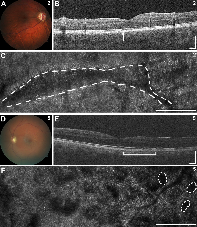
Figure 2.
Posterior segment images of selected individuals. (A–F) In individual 2, fundus photography (A) demonstrated peripapillary atrophy and linear streaks across the retina with corresponding focal disruptions of the ellipsoid zone band on OCT (B, arrow) and linear regions of abnormal photoreceptor pattern by AOSLO (C, dashed line). In individual 5, fundus photography (D) revealed subretinal deposits along the fovea with irregularities in the inner and outer segment junction on OCT (E, bracket) and patchy regions of abnormal photoreceptor pattern on AOSLO with several outlined with dashed lines (F). Scale bars: 250 µm.

