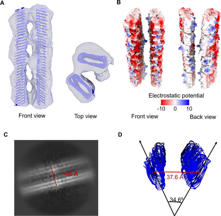
Fig 5.
Predominant spatial organization of CsgA fibrils. (A) A 2-CsgA-fibril pair (front and top views). The CsgA model (PDB: 8ENQ) was docked into the class 3 cryo-EM map generated with a 260-pixel box size. (B) Electrostatic potential of a 2-CsgA-fibril pair uncovered the hydrophobic attraction between the side of two CsgA fibrils and the electrostatic repulsion (negative to negative charge) between the front of two CsgA fibrils. (C) A representative 2D class of 2-CsgA-fibril pair (side view). (D) The fibrils were tilted 34.6° relative to each other, and the distance between their centers was 37.6 Å. The angle and distance were measured in PyMoL.

