
Figure 5.
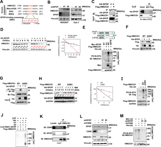
SPOP is involved in HMGCS1 dysregulation. A) Amino acid sequence alignment of putative SPOP binding consensus (SBC) motifs in HMGCS1. MacroH2A, ERG and BRD4 are known SPOP substrates containing well‐characterized SBC motifs. B) SPOP KD increased HMGCS1 steady‐state expression in 293T and Huh‐7 cell line. C) Immunoblot analysis of immunoprecipitates obtained from 293T cells transfected with indicated constructs and treated with MG132 for 6 h (left panel). HMGCS1 interacts with SPOP in vitro (right panel). Indicated constructs were transcribed and translated using TnT kit. Protein‐protein interaction was assayed by co‐IP experiments using indicated antibodies. HMGCS1 interacts with SPOP. D) Representative immunoblots showing HMGCS1 protein turnover rate in 293T cells treated with CHX, in the presence of SPOP. SPOP overexpression increased the turnover rate of HMGCS1. E) Immunoblot analysis of HMGCS1 ubiquitination from 293T cells transfected with the indicated constructs and treated with MG132 for 6 h. F) Immunoblots showing HMGCS1 steady‐state expression in indicated 293T cells transfected with HMGCS1‐ΔSBC. HMGCS1‐ΔSBC is resistant to SPOP‐mediated degradation. G) HMGCS1‐WT, but not HMGCS1‐ΔSBC, interacts with SPOP based on IP assay. H) SPOP overexpression cannot increase HMGCS1‐ΔSBC protein turnover rates in 293T cells with CHX treatment. I) HMGCS1‐ΔSBC is resistant to poly‐ubiquitination assayed by ubiquitination assay. J) Immunoblot of HMGCS1 poly‐ubiquitination in an in vitro ubiquitination assay by the CUL3‐RBX1‐SPOP E3 ligase complex. K) HMGCS1 interacts with SPOP and CSN6 in liver cancer cell. Immunoblot analysis of the indicated proteins from immunoprecipitates obtained from Huh‐7 cells treated with MG132 for 6 h. L) CSN6 KD led to reduced HMGCS1 steady‐state expression via upregulating SPOP. Immunoblot analysis of indicated proteins in 293T cells transfected with the indicated constructs. M) CSN6 KD‐mediated increased ubiquitination of HMGCS1 is SPOP‐dependent. Immunoblot analysis of poly‐ubiquitinated HMGCS1 in 293T cells transfected with the indicated constructs and treated with MG132 for 6 h.