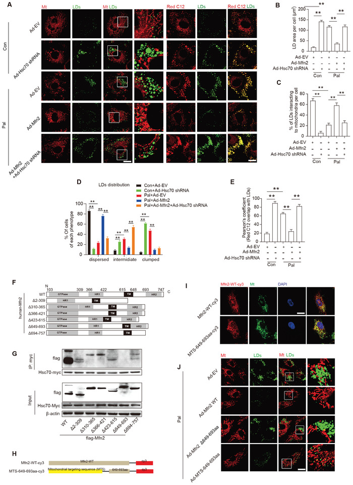
Figure 4.

Mitochondrion‐localized Mfn2 promoted MLC FAs transfer in a Hsc70‐dependent manner. A) Control‐ or Pal‐ treated primary cardiomyocytes were transfected with control adenovirus (Ad‐EV) or adenovirus expressing Mfn2 (Ad‐Mfn2) or Hsc‐70 shRNA (Ad‐Hsc70 shRNA), then LDs were labeled with Bodipy 493/503 (green), mitochondria was labeled with Mito‐tracker Red (red) and FAs were labeled with red C12 (red) respectively. Original magnification ×600. B) Mean LDs area per cell (Mean± SEM, n = 5 independent experiments, 30 cells were quantified per group, **P<0.01). C) Percentage of LDs with direct contact to mitochondria in cells treated as in A (Mean± SEM, n = 5 independent experiments, 30 cells were quantified per group, **P<0.01). D) Percentage of cells treated as in A and presenting different degrees of LD dispersion (Mean± SEM, n = 5 independent experiments, 15 images were quantified per group, **P<0.01). E) Relative localization of Red C12 with LDs was measured in cells treated as in A (Mean± SEM, n = 5 independent experiments, 30 cells were quantified per group, **P<0.01). F) Schematic diagrams of Mfn2 structure and its truncated forms. G) 293T cell were transfected with wild‐type Mfn2 and its truncated mutant and applied to immunoprecipitation assay. H) Schematic diagrams showing recombinant sequence encoding cy3 labeled wild‐type Mfn2 and mitochondrial‐targeted Mfn2 649–693aa. I) Mitochondrial localization of Mfn2‐WT‐cy3 and MTS‐649‐693aa‐cy3 were indicated by fluorescent images. J) Palmitate‐treated primary cardiomyocytes were transfected with adenovirus expressing wild‐type Mfn2 (Ad‐Mfn2 WT) or 649–693aa deletion mutant Mfn2 (Ad‐Mfn2 Δ649‐693) or MTS‐649‐693aa, then LDs were labeled with Bodipy 493/503 (green) and mitochondria was labeled with Mito‐tracker Red (red).
