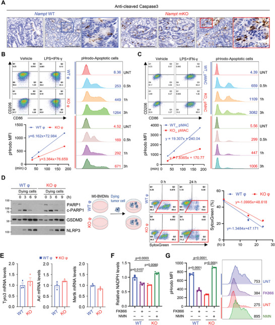
Figure 6.

NAMPT in macrophages is required for efficient clearance of apoptotic tumor cells. A) Paraffin‐embedded colonic tumor sections from AOM/DSS‐treated mice were stained with anti‐cleaved caspase3. Scale bar = 100 µm. B,C) Flow cytometry analysis of CD86 and CD206 expressions in CD11b+F4/80+ macrophages upon LPS/IFN‐γ treatment (left upper). The phagocytic activity of BMDMs (B, left bottom) and pMAC (C, left bottom), treated with LPS/IFN‐γ for 12 h was measured from 30 min to 180 min after adding dying MC38 cells labeled with pHrodo green dye using flow cytometry. Representative histogram of pHrodo intensity is shown (B and C, right).D) Western blot analysis of co‐culture experiment of BMDMs with dying MC38 cells for different time points (left). Flow cytometry analysis of SytoxGreen‐stained population during co‐culture of BMDMs and dying MC38 cells (right). E) Relative mRNA levels of Tyro3, Axl, Mertk genes in BMDMs from WT and Nampt mKO mice. F) NADPH levels are measured in WT and Nampt KO BMDMs treated with FK866 or NMN. G) BMDMs were treated with LPS/IFN‐γ for 12 h in the presence or absence of FK866 or NMN. The phagocytic activity of BMDMs was measured 2 h after adding dying MC38 cells labeled with pHrodo green dye by using flow cytometry (left). Representative histogram of pHrodo intensity is shown (right). Results are represented as the mean ± SEM. Statistical analysis was performed using the unpaired two‐tailed Student's t‐test.
