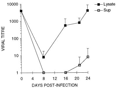
FIG. 4.
Accumulation of infectious CMV in trophoblast supernatants and cell lysates as a function of time after challenge. Villous trophoblasts from term placentas were cultured 5 days with EGF and challenged with CMV strain AD169 at an MOI of 1.0 as described in Materials and Methods. At the indicated times (horizontal axis) after challenge, 100 μl of supernatant (Sup) was removed, the adherent layer was washed with PBS, and the cells were lysed in 100 μl of medium (Lysate). Viral titer (IF/milliliter; vertical axis) was calculated from the HEL IF assay (see Materials and Methods). Each point is the mean ± SD of three replicate cultures from one of two independent experiments.

