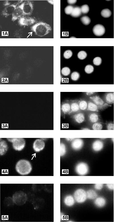
FIG. 6.
Detection and localization of kaposin protein in transformed Rat-3 cells, tumor-derived cells, and the PEL cell line BCBL-1. IFA shows cytoplasmic staining of kaposin protein in tumor-derived cell line R3/kap-TL1, using anti-kap-2 antibody (1:50) (panel 1A). Also shown are R3/kap-TL1 cells stained with preimmune serum (1:50) (panel 2A) and vector-transfected cell line R3/BK-G1 stained with anti-kap-2 antibody (1:50) (panel 3A). BCBL-1 cells stained with anti-kap-2 antibody (1:5) and preimmune serum (1:5) are shown in panels 4A and 5A, respectively. Intense staining with anti-kap-2 antibody in a restricted region of the cytoplasm of R3/kap-TL1 and BCBL-1 cells are indicated by arrows in panels 1A and 4A, respectively. DAPI staining of nuclei of the above-specified fields are shown in panels 1B, 2B, 3B, 4B, and 5B, respectively. Magnifications: panels 1 to 3 (A and B), ×1,800; panels 4 and 5 (A and B), ×3,000.

