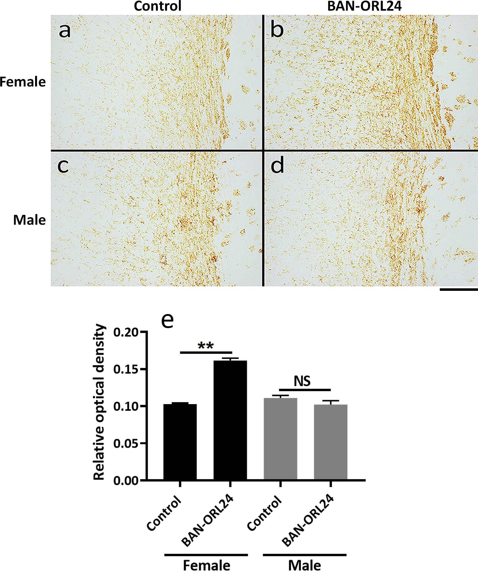
FIGURE 10. The corpus callosum of NOR antagonist-treated female pups exhibits increased myelination.

Nine-day-old female and male pups were daily administered the NOR antagonist BAN-ORL24, as indicated in the legend for Figure 9. Animals were sacrificed at postnatal day 14 and myelination of the corpus callosum was assessed after immunohistochemical staining of coronal brain sections with MBP antibody. Shown are representative images corresponding to comparable corpus callosum regions for the female (a and b) and male (c and d) pups. Scale bar: 200 μm. MBP immunostaining of the corpus callosum was quantified using the NIH ImageJ program for the assessment of DAB staining as indicated under “Methods”. ** Control vs BAN-ORL24 treated females, p<0.01.
