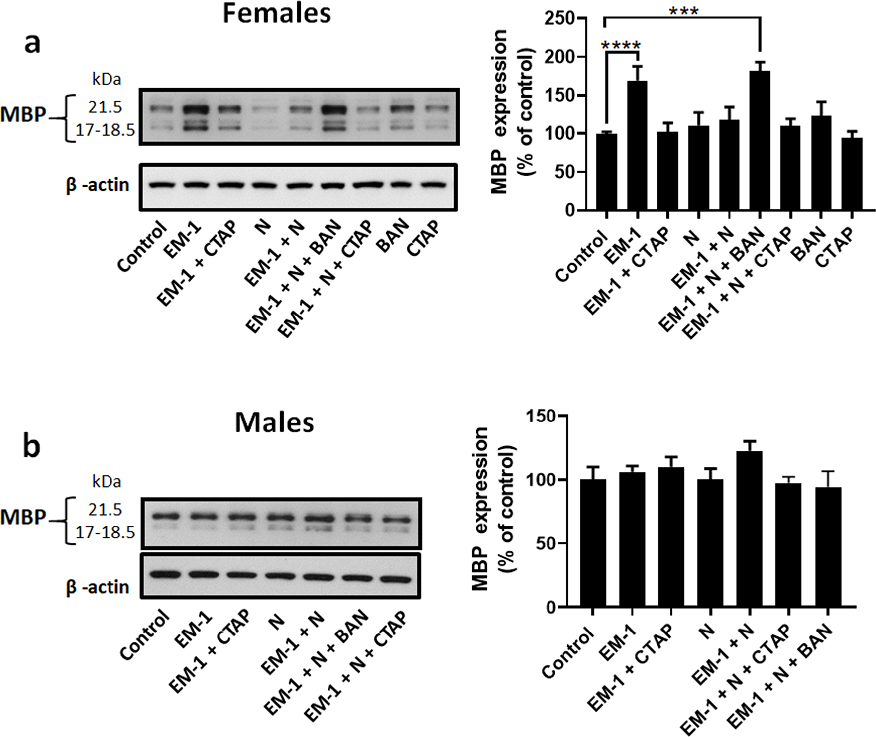
Figure 3. Treatment of pre-oligodendrocytes with EM-1 and nociceptin induces sex-specific effects on MBP expression.

Developing oligodendrocytes from 9-day-old rat brain were cultured for 72 hours in chemically defined medium (CDM) alone (control) or in CDM supplemented with 1 μM Endomorphin-1 (EM-1), 1 μM EM-1 + 1 μM CTAP (EM-1 + CTAP), 1 μM Nociceptin (N), 1 μM EM-1 + 1 μM N (EM-1+ N), 1 μM EM-1 + 1 μM N + 100 nM BAN-ORL24 (EM-1 + N + BAN), 1 μM EM-1 + 1 μM N + 1 μM CTAP (EM-1 + N + CTAP), 100 nM BAN-ORL24or 1μM CTAP (CTAP). MBP expression levels were determined by western blot analysis using β-actin as loading control. Shown are representative western blots for cell cultures prepared from (a) female and (b) male pre-oligodendrocytes. Results in the bar graphs are expressed as % of the control values and represent the mean ± SEM from at least 3 separate experiments carried out in at least duplicates. ***p<0.0005, ****p<0.0001.
