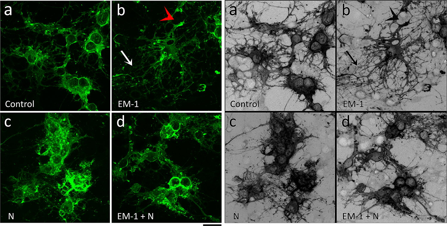
FIGURE 4. EM-1 stimulates the morphological maturation of female oligodendrocytes and this effect is abolished by nociceptin.

Developing OLGs from 9-day-old female rat brain were cultured for 72 hours in chemically defined medium (CDM) alone (Control) or supplemented with 1 μM EM-1, 1 μM N, or 1 μM EM-1 + 1 μM N. Cells were stained with anti-MBP antibody and visualized by confocal microscopy. Representative images demonstrate that in comparison with CDM alone (a), EM-1 (b) stimulates the formation of membrane extensions (red arrow) and the branching and length of processes (white arrow). This effect is however abolished by co-incubation with nociceptin (d). No apparent treatment effects were observed for nociceptin alone (c). Scale bar: 20 μm.
