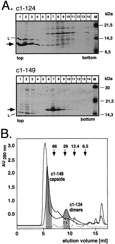
FIG. 4.
Quaternary structure of core protein c1–124. (A) Sedimentation analysis. E. coli-derived protein c1–124 (upper panel) was sedimented on an analytical sucrose gradient. Aliquots from each fraction were analyzed by SDS-PAGE and staining with Coomassie blue. Essentially all of the protein was present in the four top fractions (arrow), cosedimenting with the lysozyme (L) used during preparation of the cell lysate. The molecular masses of the marker proteins (lane M) are indicated on the right. Protein c1–149 was analyzed in parallel (lower panel) and was present mainly in fractions 6 to 8. (B) Size exclusion chromatography. Bacterial lysates containing protein c1–124 or c1–149 were analyzed on a Superdex 75 column equilibrated in PBS. The elution profiles and the positions of the two proteins as determined by SDS-PAGE are indicated. The dashed line shows the elution profile for a set of protein markers with the indicated molecular masses. AU280 nm, absorbance units at 280 nm.

