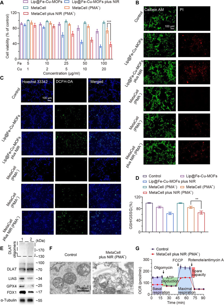
Fig. 4. In vitro anticancer effects of MetaCell on 4T1 cancer cells.
(A) Anticancer effects of MetaCell on 4T1 cells. Cancer cells were coincubated with either Lip@Fe-Cu-MOFs or MetaCell with or without PMA. After a 4-hour incubation, the cells were treated with NIR and then evaluated with the MTT assay 24 hours later. (B) Live-dead staining of 4T1 cells after the treatment. 4T1 cells were coincubated with Lip@Fe-Cu-MOFs or MetaCell with or without PMA and then irradiated with NIR light. After irradiation, cells were stained with calcein AM–PI dye to visualize living and dead cells. (C) Intracellular ROS levels after the treatment. The effect of MetaCell treatment on the ROS generation was measured using the ROS dye DCFH-DA. (D) The intracellular levels of GSH in 4T1 cells after the MetaCell treatment, measured with a GSH/GSSG ratio assay kit. (E) Western blot analysis of the protein expressions of DLAT, LIAS, FDX1, and GPX4 in 4T1 cells after treatment with either neutrophils (1) or MetaCell (2) with PMA and NIR irradiation. (F) An ultrastructural analysis of MetaCell-treated 4T1 cells using TEM. (G) The oxygen consumption rate (OCR) of 4T1 cells after the treatment with either NEs or MetaCell with PMA and NIR. This provides insights into the impacts of the treatments on cellular respiration. The data in all panels are presented as mean ± SD (n = 3). **P < 0.01 and ***P < 0.001 (Student’s t test, two tails).

